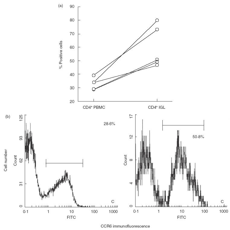
Fig. 3.

Flow cytometric analysis for CCR6 from patients’ CD4+ IGL and CD4+ PBMC. The same patient’s IGL and PBMC were analysed for the expression of CCR6. Compensation was made for each colour with positively and negatively stained control cells. Lymphocytes were gated according to their side scatter properties, and 104 cells in the gate were monitored for two-colour analysis. Isotype-matched antobodies were used to define the background threshold. (a) Percentage of CCR6-positive cells in CD4+ IGL and CD4+ PBMC of five patients with periodontal disease. (b) Flow cytometry results showing CCR6 expression on CD4+ IGL (right panel) and CD4+ PBMC (left panel) of one patient with periodontal disease.
