
Fig. 6.
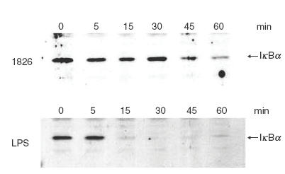
Kinetics of IκBα degradation in the CpG ODN 1826 activated macrophages. RAW 264·7 macrophages (1 × 10⁶ cells/well) were activated with 1μg/ml of CpG ODN 1826 (a) or 10 ng/ml of LPS for different time intervals before the cells were lysed in lysis buffer and analysed for IκBα by immunoblotting.