
Fig.1.
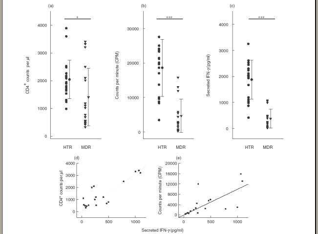
Lymphoproliferative responses, CD4 counts and IFN-γ production in PBMC from patients with MDRTB and HTR in response to the PPD antigen of Mycobacterium tuberculosis. (a) PBMC were isolated and FACS analysis performed. CD4 count was calculated based on a concomitant complete blood count. (b) PBMC were stimulated for 5 days with the PPD antigen of M. tuberculosis at a concentration of 1·0 μg/ml. Proliferative responses were assessed as 3H]-thymidine incorporation in PBMC from healthy controls and TB patients. Incorporation of 3H]-thymidine occurred during the last 18 h of a 5 day culture; unstimulated PBMC served as controls. (c) PBMC were stimulated for 96 h with PPD antigen at a concentration of 1·0 μg/ml. Supernatant fluids were prepared following a 96 h stimulation with PPD, and IFN-γ production was measured using ELISA. Values are the mean ± s.d. of triplicate supernatant samples. *P < 0·05; **P < 0·01; ***P < 0·001 (Student’s t-test). Significant correlations were found between (d) IFN-γ and CD4 counts (n = 18, r = 0·85, P < 0·001), and (e) between IFN-γ and lymphoproliferative responses in TB patients (n = 18, r = 0·68, P < 0·01).