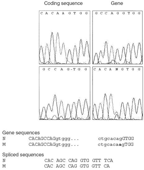
Fig. 3.

Characterization of the mutation. Electropherograms of the mutated region in the coding and gene sequences of patient 2 and one parent are shown. At the position of the mutation in intron IX, two overlapping bases can be read in the electropherogram from the parent (N, for G and A), while a missing base in the coding sequence from the patient is indicated by a –. Nucleotide sequences of the gene and coding regions of the normal (N) and mutated (M) alleles are given. The mutation is indicated in bold, and exon nucleotides are represented in block characters.
