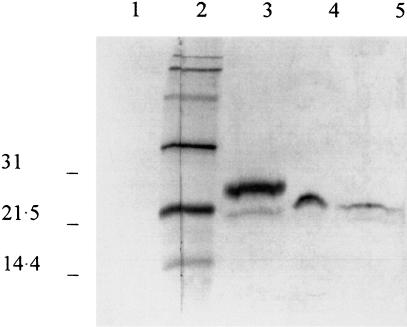
Fig. 1.
Western-blot analysis of IL-1ra protein in lysate and supernatant of oral mucosal cells. Lane 4 is a fraction of a concentrated pool of supernatants from three unstimulated cultures. Cell-associated IL-1ra is shown from unstimulated cells (lane 5). For comparison, lane 3 is the 25 kD monocyte IL-1ra. Lane 2 is the biotinylated standard (mol. wt. × 103 daltons). Lane 1 is the control with preimmune serum.

