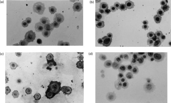
Fig. 2.
Immunohistological staining in scattered oral mucosal cells (×20). These results are representative of two similar experiments. (a) IL-1ra staining is observed in a few unstimulated cells. (b) It is more intense in the cytoplasm of cells stimulated with TGF-β1. (c) These cells stain markedly positive for involucrin. (d) Staining was nil in control with preimmune serum. Control was also negative with anti-IL-1ra serum preincubated with recombinant IL-1ra.

