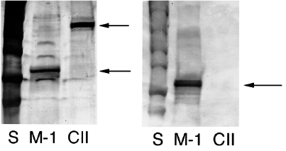
Fig. 2.
Western blot and silverstaining. (a) Silverstaining and (b) Western blot (reduced conditions) of the protein batches of CII and matrilin-1 that were used. S, standard; m-1, matrilin-1; CII, collagen type II. Arrows indicating positive signals from m-1 and CII, showing that no m-1 was found in the CII preparation.

