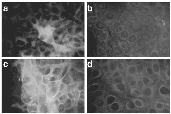
Fig. 2.

Immunofluorescence staining of HAV antigen (a and b) and MHC class I (c and d) expressed in Hu-H7 cells. Hu-H7 cells were transfected with full-length (a and c) or truncated, ΔX (b and d) HAV ssRNA created by in vitro transcription. Immunofluorescence staining was performed 48 h after transfection.
