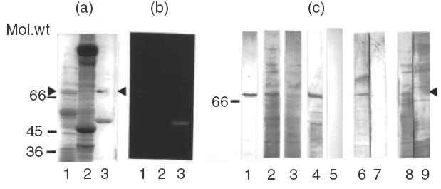
Fig. 1.

Correlation of SucDH-Fp fluorescence and immunoreaction with myocarditis serum. (a) SDS-PAGE followed by Coomassie blue staining of: lane 1, rat liver inner mitochondrial membrane proteins (10 μg); lane 2, bovine heart mitochondrial proteins (30 μg); lane 3, 6HDNO (5 μg); the upper band in the lane marks the position of the fluorescent protein band shown in (b) (arrowhead). Molecular weight markers are indicated on the left hand site of the panel. (b) Fluorescent protein bands revealed on the gel shown in (a) by a UV-transilluminator following incubation in 10% acetic acid. Lanes same as in (a). (c) Rat liver inner mitochondrial membrane proteins (lanes 1 and 2) and bovine heart mitochondrial proteins (lane 3) were separated by SDS-PAGE, blotted to nitrocellulose membrane and individual lanes of the Western blot were developed with: lane 1, rabbit α-6HDNO antiserum; lane 2, myocarditis serum; lane 3, myocarditis serum; lane 4, the fluorescent protein band shown in (b), lane 1, was excised, eluted from the gel, applied to the same gel used to separate the mitochondrial proteins and the corresponding lane of the Western blot was developed with myocarditis serum; lane 5, Western blot of inner mitochondrial membrane proteins reacted with human control serum; lanes 6, 7, and 8, 9 present Western blots performed with rat liver inner mitochondrial membrane proteins, developed either with rabbit α-6HDNO antiserum before (lane 6) and after preincubation with 100 μm FAD for 1 h at room temperature (lane 7), or with myocarditis serum before (lane 8) and after preincubation with FAD (lane 9). Sera without added FAD were incubated for the same time with FAD at room temperature for 1h.
