
Fig. 3.
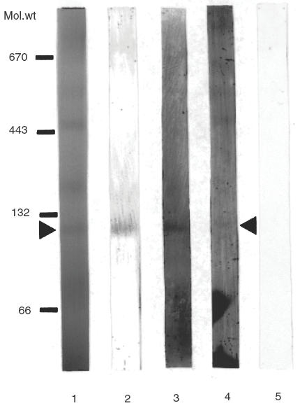
Separation of inner mitochondrial membrane protein complexes by BN-PAGE followed by Western blotting. Rat liver inner mitochondrial membrane protein complexes were solubilized with digitonin and separated on a 6% to 16·5% gradient polyacrylamide gel (lane 1). Following electrophoresis, the gel was blotted to PVDF membrane, and individual lanes were cut and developed with mouse monoclonal antibody against SucDH-Fp (lane 2), with rabbit α-6HDNO antiserum (lane 3), with myocardatis serum (lane 4) and with human control serum (lane 5). Position of molecular weight markers are indicated on the left hand side of the figure (132/66 kDa, bovine serum albumin dimer and monomer, respectively; 443 kDa, horse spleen apoferritin; 670 kDa bovine thyroglobulin).