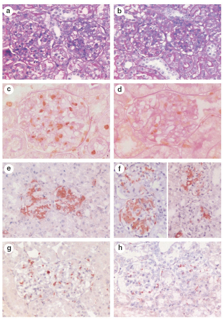
Fig. 2.

Glomerular histology in control treated rats with mesangial proliferative GN (a, c, e, g) and IL-10 treated rats with GN (b, d, f, h). Glomeruli from control rats with GN showed proliferative GN (a), the severity of which was reduced in IL-10 treated rats (b). Proliferating cells were plentiful in control rats with GN, and could be identified by immunohistochemistry for PCNA (c, brown reaction product). The number of PCNA + cells was reduced by IL-10 treatment (d, brown reaction product). Glomerular α-smooth muscle actin expression was not reduced significantly by IL-10 treatment (Ctrl GN, e and IL-10 GN, f). Glomerular ED1 + macrophages were reduced by IL-10 treatment (Ctrl GN, g and IL-10 GN, h, brown reaction product). Panels (a) and (b) were stained with PAS. (c–h) Immunoperoxidase with DAB substrate and PAS counterstain in panels (c) and (d), haematoxylin counterstain in (e–h). Magnifications: a, b, g, h and e × 300, f× 350, c × 500, d × 600.
