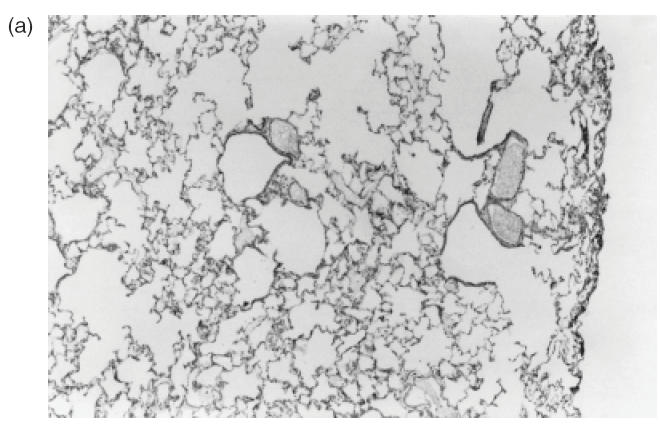
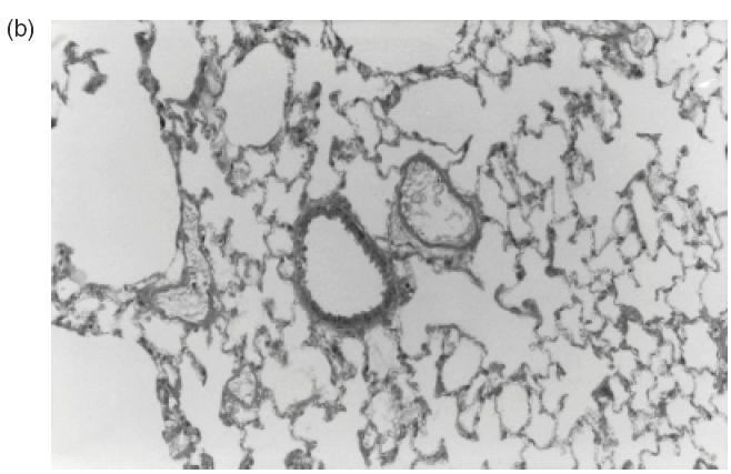
Fig. 3.


Representative sections of pulmonary tissue of Wistar rats after 24h of the intrajugular injection of normal human IgG. (a,b) Haematoxylin and eosin-stained cryosections. Lack of perivascular cellular infiltrate in lung tissue was detected in all animals injected with normal IgG (original magnification ×20 in (a) and ×40 in (b)).
