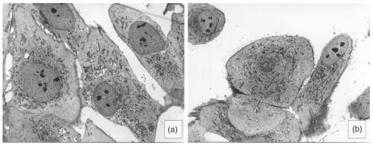
Fig. 2.

Ultrastructure of T-HPMC in comparison to P-HPMC. By transmission electron microscopy T-HPMC (a, ×725) and P-HPMC (b, × 725) display nuclei and nucleoli with similar sizes and shapes and cytoplasms with similar tinctorial patterns, although microvilli are abundant in P-HPMC and few in T-HPMC.
