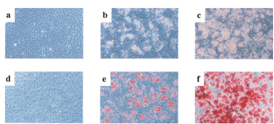
Fig. 5.

Troglitazone-mediated differentiation of FLS into adipocyte-like cells. FLS were cultured with or without troglitazone for three weeks. a-c: Phase contrast microscopy, demonstrating morphological changes in FLS. (a) unstimulated FLS; (b) FLS cultured with 1 μm of troglitazone. c: FLS cultured with 10 μm of troglitazone. Original magnification, 200×. The morphological changes included transformation to round cells and the appearance of lipid-like vesicles in the cytoplasm. These changes were troglitazone dose-dependent. (d–f) Detection of lipid accumulation in FLS by Oil Red O staining. (d) unstimulated FLS; (e) FLS cultured with 1 μm of troglitazone; (f) FLS cultured with 10 μm of troglitazone. Original magnification, ×200. Note that lipid accumulation in FLS was troglitazone dose-dependent. Results shown are representative data of five experiments.
