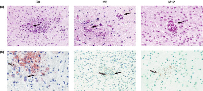
Fig. 3.
Representative sections of the brains of SCID mice, humanized with PBMC from HIV-infected patients after treatment with HAART for various periods of time, infected with T. gondii. (a) haematoxylin-eosin staining for cyst detection and (b) antiperoxidase moAb staining for tachyzoite detection. Brain parenchyma from a mouse humanized with PBMC from an HIV-infected patient before HAART (D0) shows necrotic foci, a large number of cysts (+ + + +) and numerous tachyzoites. Brain parenchyma from a mouse humanized with PBMC from an HIV-infected patient after 6 months of HAART (M6) shows necrotic foci, with a few cysts (+ to + +) and a few tachyzoites. Brain parenchyma from a mouse humanized with PBMC from an HIV-infected individual after 12 months of HAART (M12). The section shows moderate necrotic foci with a few cysts (+ + to + + +) and numerous tachyzoites. Original magnification: ×400

