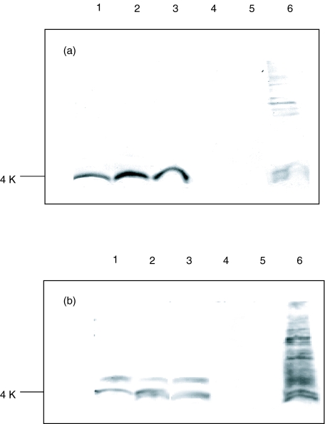
Fig. 5.
Detection of Aβ peptides [1–40], [1–42] and [1–43] and total Aβ in FAD brain by 1E8-4b (a) and 1E8 (b) antibodies using immunoblotting followed by ECL development. Samples were electrophoresed on 14% Tris-tricine gels with 100 ng/well of Aβ peptides [1–40], [1–42] and [1–43] and APP peptides 695 and 770. A total of 5 μl of FAD brain sample was used. The recombinant Fab 1E8-4b and the isotype control M2 anti-Flag monoclonal antibody were applied to the immunoblots at 5 μg/ml, while 1E8 was used at 50 μg/ml. The isotype control displayed no reactivity to the peptides or FAD brain sample. Lane 1: Aβ peptide [1–40]; lane 2: Aβ peptide [1–42]; lane 3: Aβ peptide [1–43]; lane 4: APP peptide 695; lane 5: APP peptide 770; lane 6: FAD (PS-1mut) brain sample.

