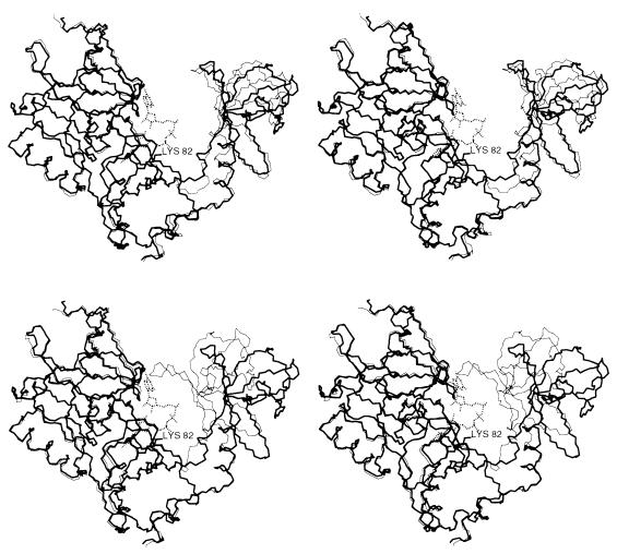
Figure 1.
Comparison between the structure of capping enzyme (thick line) complexed with cap analogue (broken line) and the previously determined structures (thin lines) of GTP complexes in open (a) and closed (b) conformation. Only the main chain and the Lys-82 side chain are shown. (a) The upper part of domain 2 is approximately 4 Å closer to the viewer than in the open GTP complex, but the size of the cleft between the domains is similar. (b) The closed GTP complex conformation is sterically incompatible with the presence of the cap analogue in the active site. In each case, the Cα atoms of residues 11–230 of domain 1 were superimposed.

