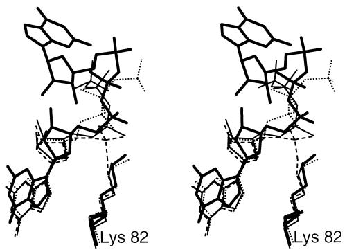
Figure 3.
Comparison of ligands bound in the active site of PBCV-1 capping enzyme. Closed GTP complex (thin line), open GTP complex (thin, dotted line), and guanylylated adduct (thin, broken line) were superimposed on the cap analogue complex (thick line) by using the Cα atoms of residues 11–230 of domain 1.

