
Fig. 1.
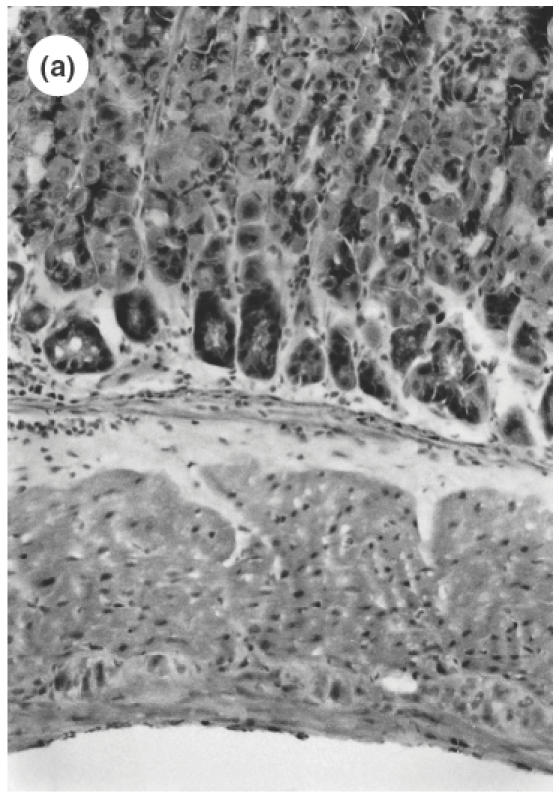
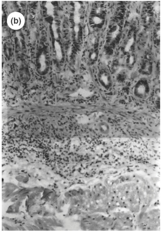
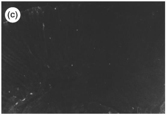
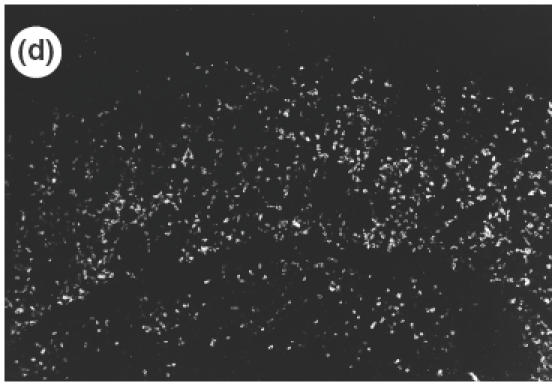
Microscopic pictures of the gastric mucosa of SS1-infected mice compared with noninfected control mice. (a) Control mice (H&E staining, ×100). (b) SS1-infected mice (H&E staining, ×100). (c) Immunohistochemical study of CD4 positive cells in the stomach of control mice. (d) Immunohistochemical study of CD4-positive cells in the stomach of SS1-infected mice. The same specimens as (a) and (b) were observed.