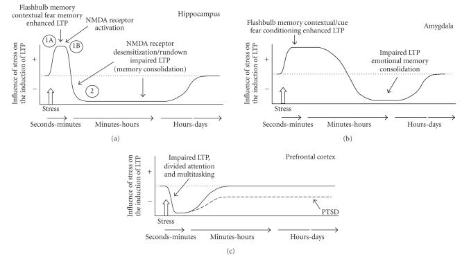
Figure 3.
Temporal dynamics model of how stress affects memory-related processing in the hippocampus, amygdale, and prefrontal cortex. The initiation of a strong emotional experience activates memory-related neuroplasticity in the hippocampus and amygdala, and suppresses PFC functioning (phase 1). The most rapid actions would involve increases in ACTH, CRF, NE, acetylcholine, dopamine, and changes in GABA receptor binding (phase 1A), followed within minutes by elevated levels of glucocorticoids (phase 1B). The combination of the activation of the hippocampus by these neuromodulators with coincident tetanizing stimulation produces a great enhancement of LTP. Within minutes of the initiation of phase 1, the hippocampus undergoes a reversal of its plasticity state, based, in part, on the reduction in the sensitivity of NMDA receptors (phase 2). Tetanizing stimulation delivered to the hippocampus during phase 2 will thereby result in an impairment of the induction of LTP. The amygdala continues in its form of phase 1 longer than the hippocampus, but eventually, the amygdala, as well, exhibits an inhibitory phase, perhaps as it is involved in the consolidation of the emotional memory. The PFC is only inhibited by stress; the recovery from its suppression of functioning would depend on the nature and intensity of the stressor, interacting with the ability of the individual to cope with the experience. In the case of trauma-induced PTSD, the PFC may not recover to its original state of efficiency in suppressing the activity of lower brain areas, such as the amygdala and brain stem nuclei.

