
Figure 7.
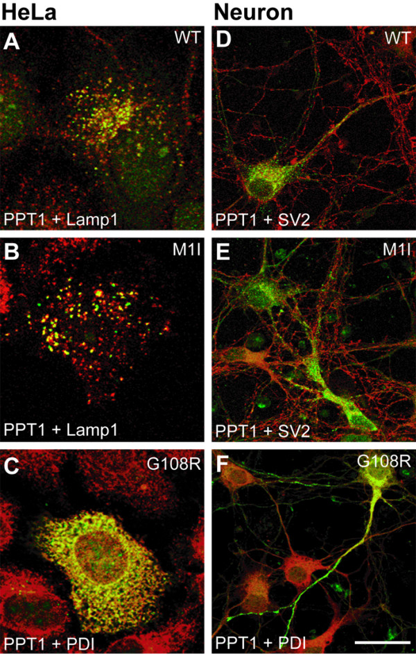
Intracellular localization of wild type and mutant PPT1 in non-neuronal and neuronal cells. HeLa cells (A-C) transfected with pCMV5-PPT1 plasmids and neurons (D-F) infected with PPT1-SFV bearing the indicated mutations were double-stained for PPT1 using the GST-PPT1 antibody (green), for lysosomes using the Lamp-1 antibody (red), for synaptic vesicles using the SV2 antibody (red) and for ER using the PDI antibody (red). Colocalization is shown in yellow. Scale bar = 20 μm.