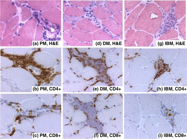
Figure 2.

Perivascular localization of CD4+ and CD8+ T lymphocytes. Immunohistochemical staining of samples from patients with polymyositis (PM) (a-c), dermatomyositis (DM) (d-f), and inclusion-body myositis (IBM) (g-i). (a) Hematoxylin and eosin (H&E) staining to localize inflammatory cell infiltrates in a patient with polymyositis. (b) CD4+ T lymphocytes stained with a monoclonal SK3 mouse IgG1 antibody in the same area as in (a) but further down in the biopsy. (c) CD8+ T lymphocytes stained with a monoclonal SK1 mouse IgG1 antibody in a consecutive section to that in (b). (d) H&E staining to localize inflammatory cell infiltrates in a patient with dermatomyositis. (e) CD4+ T lymphocytes stained with a monoclonal SK3 mouse IgG1 antibody in the same area as in (d) but further down in the biopsy. (f) CD8+ T lymphocytes stained with a monoclonal SK1 mouse IgG1 antibody in a consecutive section to that in (e). (g) H&E staining to localize inflammatory cell infiltrates in a patient with inclusion-body myositis. (h) CD4+ T lymphocytes stained with a monoclonal SK3 mouse IgG1 antibody in the same area as in (g) but further down in the biopsy. (i) CD8+ T lymphocytes stained with a monoclonal SK1 mouse IgG1 antibody in a consecutive section to that in (h). Original magnifications: ×312.5 (a-c, g-i) and ×250 (d-f).
