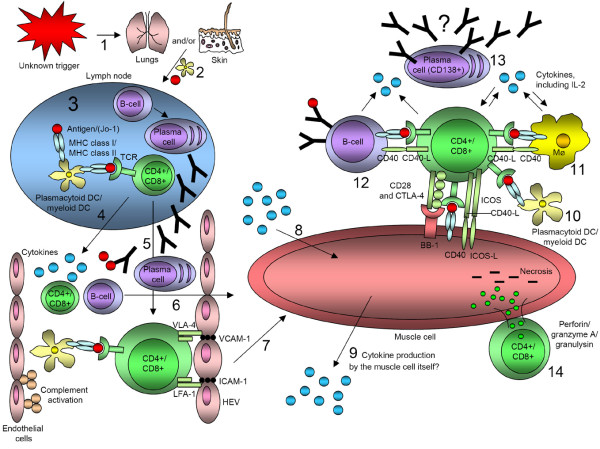
Figure 3.

Hypothetical involvement of T lymphocytes, B lymphocytes, and dendritic cells (DCs) in idiopathic inflammatory myopathies. (1) An unknown trigger (for example viral infection or ultraviolet radiation) in the respiratory tract or through the skin leads to the cleavage of histidyl-tRNA synthetase by granzyme B through antiviral CD8+ T lymphocytes in the lungs. (2) Immature DCs carry receptors on its surface that recognize common features of many pathogens. When a DC takes up a pathogen in infected tissue it becomes activated and migrates to the lymph node. (3) Upon activation, the DC matures into a highly effective antigen-presenting cell (APC) and undergoes changes that enable it to activate pathogen-specific lymphocytes in the lymph node. T lymphocytes become activated and B lymphocytes, with active help from CD4+ T lymphocytes, proliferate and differentiate into plasma cells. (4) Activated DCs, T lymphocytes, and B lymphocytes could release cytokines into the bloodstream. (5) The activated T lymphocyte, on which the DC-MHC-antigen complex is bound, itself binds to specialized endothelial cells called high endothelial venules (HEV). For this purpose it uses the VLA-4 (very late activation antigen-4) and LFA-1 (lymphocyte function associated antigen-1) molecules on its surface to interact with adhesion molecules (vascular cell-adhesion molecule-1 (VCAM-1) and intercellular cell-adhesion molecule-1 (ICAM-1)) on HEVs, where they can penetrate into peripheral lymphoid tissues. (6,7) Naïve T lymphocytes and B lymphocytes that have not yet encountered their specific antigen circulate continuously from the blood into the peripheral lymphoid tissues. (8,9) Various cytokines from the bloodstream or produced locally could affect the muscle tissue or cell in many different ways. However, it is not clear whether the muscle cell itself could produce and release cytokines. (10–12) DCs, macrophages (Mϕ), and B lymphocytes can interact with T lymphocytes in various ways. T lymphocytes could possibly also bind to muscle cells through inducible co-stimulators (ICOS), CD40 ligand (CD40-L), CD28, and CTLA-4 (CD152) on T lymphocytes to ICOS ligand (ICOS-L), CD40, and BB-1 antigen on the muscle cell. In that fashion, the muscle cell would function as an APC. (13) Plasma cells (CD138+) can be found in the muscle tissue of certain subgroups of patients with idiopathic inflammatory myopathy, but whether these cells could produce autoantibodies locally is not yet known. (14) T lymphocytes have been shown to bind in close contact with muscle cells and to release perforin, granzyme A, and granulysin, which may cause necrosis of muscle tissue or cells.
