
Figure 1.
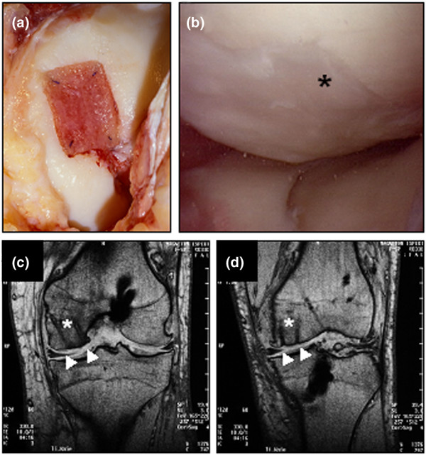
Arthroscopic and magnetic resonance imaging evaluation of cartilage defects treated with autologous chondrocyte grafts (BioSeed®-C). (a) Intra-operative situation of a cartilage defect situated at the femoral condyle covered with transosseously fixed BioSeed-C (20 mm × 30 mm). Note that the healthy cartilage rim is partly intact. (b) At 9 months after surgery, second-look arthroscopy showed the formation of a cartilage repair tissue of a tough condition (asterisk). Magnetic resonance imaging (MRI) at 6 months (c) and 12 months (d) after implantation of BioSeed-C shows transosseous drilling holes (white asterisks) due to fixation of the graft. The repair tissue covers the defect (white triangles) and gives a slightly altered MRI signal.