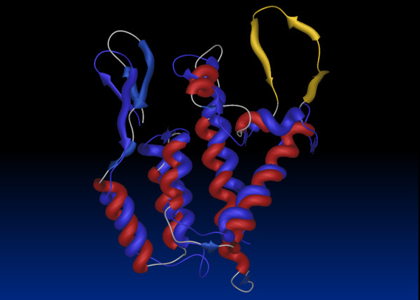
Figure 4.

Similarity between the structures of retroviral capsids. Superimposition of the structures of the N terminal domains of HIV-1 (Red) and MLV (blue) capsids demonstrates overall structural conservation although the Cyclophilin A binding loop (yellow) is absent in MLV. The pdb files for HIV-1 (1M9C) [94] and MLV (1UK7) [40] were superimposed using pairwise structure comparison [95].
