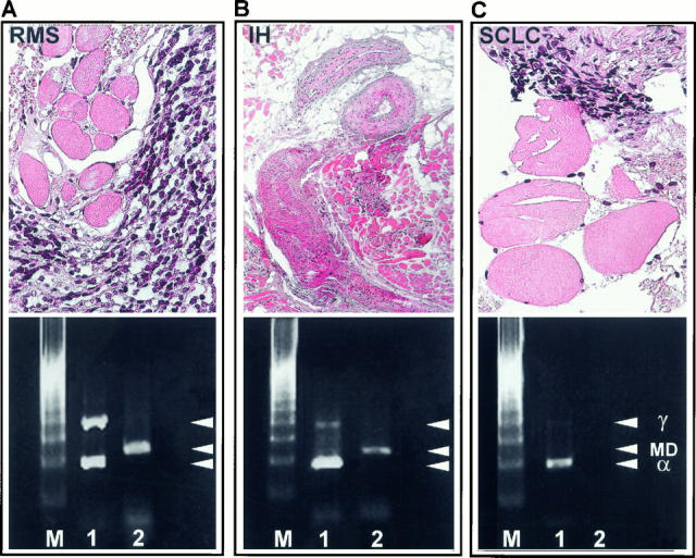
Figure 8.

Determination of the specificity of the MyoD1 RT-PCR compared to the α/γAChR duplex RT-PCR. Ex vivo embryonal RMS biopsy (case 25923/95) contaminated by skeletal muscle (A) showed strong transcription of MyoD1 and a RMS specific α/γ ratio of 0.93 (A, Lane 1), indicating a dramatic overexpression of the γAChR in RMS. In an intramuscular hemangioma (IH, case 26777/98) (B> the α/γ ratio was 6.2 (B, Lane 1) and in a biopsy derived from a small-cell lung cancer (SCLC, case 1007/99) contaminated by normal muscle (C>, the α/γ ratio was 7.3 (C, Lane 1>. By contrast, MyoD1 was detected in the intramuscular hemangioma (B, Lane 2> in quantities similar to those shown in RMS (A>, but MyoD1 mRNA was not found in the biopsy with small amounts of contaminating muscle due to its low sensitivity (C, Lane 2>. M, marker; Lane 1 = α/γAChR RT-PCR; Lane 2 = MyoD1 RT-PCR; γ = γAChR; MD = MyoD1; α = αAChR.
