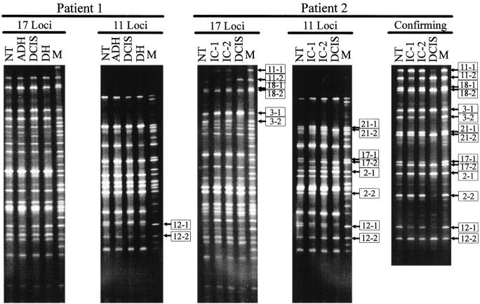
Figure 3.
Multiplex genotype determination at 28 marker loci (listed in Table 1 ) for the lesions microdissected from breast tissues of the two patients. NT, normal tissue; DH, ductal hyperplasia; ADH, atypical ductal hyperplasia; DCIS, ductal carcinoma in situ; IC, invasive carcinoma; M, mixture of PCR products separately amplified from the 28 loci in corresponding heterozygous DNA samples used as molecular markers. Bands representing different alleles are distinguished by “−1” and “−2.” Alleles for the loci with LOH are indicated by boxed numbers. The confirming panel is for confirmation of the results from the loci showing LOH in patient 2. The loci in this panel were separately amplified by using aliquots from the first-round PCR products. The PCR products from different loci for each samples were mixed, respectively, before being loaded onto the DGGE gel. Only results from tumor 2 are shown.

