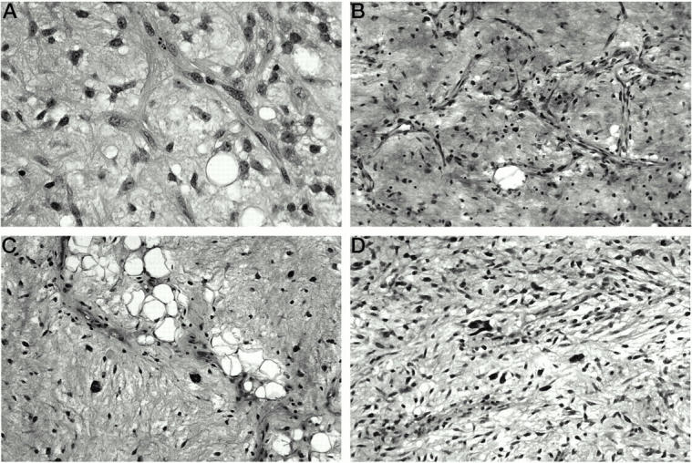
Figure 1.

A: Classic myxoid LS, low grade, with small, uniform tumor cells in a background of myxoid stroma and delicate “chicken-wire” type vascular network. Of note is the absence of the pleomorphic giant tumor cells characteristic of WDLS with myxoid changes. B: WDLS, predominantly myxoid. Although the myxoid background and the branching vasculature might suggest the diagnosis of classic myxoid LS, the tumor cells are larger and less uniform. C: WDLS, predominantly myxoid, with focal areas of lipoma-like LS and scattered tumor giant cells. D: Myxofibrosarcoma, low grade. Most of the tumor cells are uniform, but predominantly spindly, in contrast with the round to oval appearance of myxoid LS. Rare pleomorphic tumor cells are scattered within the myxoid stroma, which helps in distinguishing this tumor from classic myxoid LS.
