
Figure 1.
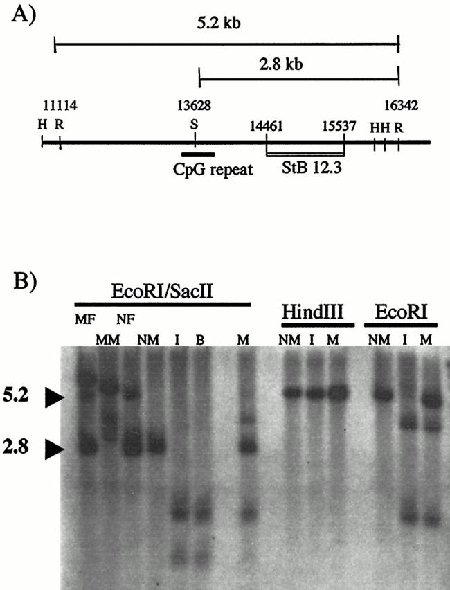
Southern analysis of index patient. A: Patient DNA is digested with EcoRI (R) and SacII (S), then probed with the cloned fragment StB12.3. Normal males generate a 2.8-kb SacII/EcoRI fragment, whereas normal females generate an additional 5.2-kb EcoRI/EcoRI fragment from the methylated allele. Additional restriction sites used in this paper (HindIII (H)) are shown. B: Lanes 1–4 show the patterns seen after digestion with EcoRI/SacII in control patients with full mutations (MM, mutant male; MF, mutant female) as compared to normal controls (NM, normal male; NF, normal female). Both the index patient (I) and his brother (B) show a loss of this normal pattern and the appearance of smaller bands. The mother (M) has a more complex pattern. Digestion with HindIII produces identical bands in a normal male, the index patient, and his mother. In contrast, digestion with EcoRI alone demonstrates the presence of a new EcoRI site in both the index patient and his mother, when compared to the normal male control.