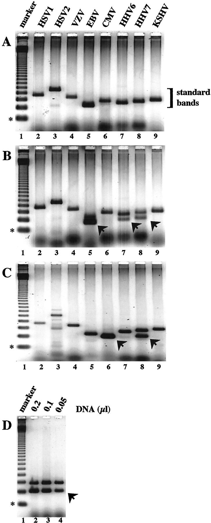
Figure 1.

Quantitative ICS-PCR analysis of eight human herpesviruses in patient samples. PCR reactions containing purified DNA derived from 10 μl whole blood and 20 molecules of the HHVQ-1 ICS standard were amplified as described in Materials and Methods. DNA isolated from three different patients (A, B, and C) was amplified with primers specific for HSV-1 (lanes 2), HSV-2 (lanes 3), VZV (lanes 4), EBV (lanes 5), CMV (lanes 6), HHV-6 (lanes 7), HHV-7 (lanes 8), and KSHV (lanes 9). The sizes of the predicted PCR products derived from the HHVQ-1 standard and the specific viruses are given in Table 1 . PCR products derived from the HHVQ-1 ICS using the virus-specific primers are bracketed as standard bands. Arrowheads indicate PCR products derived from the viral targets. The weak bands observed with the HSV-2 primers in A and C, lanes 3 are nonspecific. D: DNA from the patient analyzed in C was re-analyzed by amplification with CMV-specific PCR primers and the amount of whole blood DNA indicated. In each panel, lane 1 contains a 123-bp DNA ladder as a size marker; the 123-bp fragment is indicated with an asterisk.
