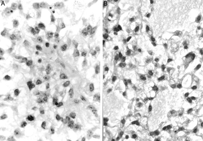
Figure 6.

Histopathological findings of biopsy specimens of KS682. A: Round mesenchymal cells show undifferentiated features with prominent nucleoli (hematoxylin and eosin, original magnification, ×780). B: Cystic change with myxoid material is seen in several foci, and some cells show lipoblastic features with vacuoles in the cytoplasm (hematoxylin and eosin, original magnification, ×780).
