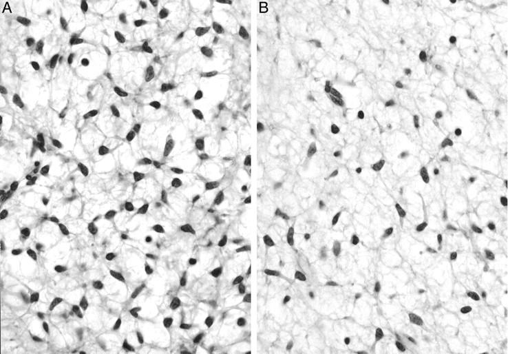
Figure 8.

Histopathological findings of biopsy specimens of KS685. A: Small spindle cells with a primitive mesenchymal appearance proliferate diffusely in an edematous background. Dendritically elongated cytoplasms form a network-like structure making vacuolar space in the myxoid stroma. Unequivocal lipoblasts, however, are not identified (hematoxylin and eosin, original magnification, ×780). B: Myxoid change in the stroma is prominent. Plexiform fine vascular network, however, is not identified (hematoxylin and eosin, original magnification, ×780).
