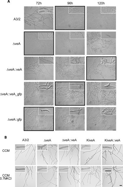
FIG. 7.
Microscopic investigations of A. chrysogenum strains as indicated. (A) For arthrospore formation, all strains were grown at 27°C and 180 rpm in liquid CCM. At the assigned time points, mycelial morphology was analyzed by differential interference contrast microscopy, and representative microscopic fields are depicted. Insets show an enlargement of characteristic mycelial structures, and black frames indicate the beginning of arthrospore formation. (B) Growth of the recipient strain and transformants in complete medium with (CCM plus 0.7 M KCl) and without (CCM) the addition of the osmotic stabilizer KCl. Insets show an enlargement of characteristic mycelial tips. The scale bar represents 20 μm in all panels.

