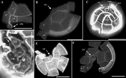
FIG. 2.
Epifluorescence microscopy of Calcofluor-stained cultured vegetative cells of A. minutum isolated from Irish coastal waters. (A) Ventral view showing the first apical plate (1′) and a narrow sixth precingular plate (6′). (B) Apical view showing a wide sulcal anterior plate (sa) and a narrow connection between the apical pore (po) and the first apical plate (1′). (C and D) Details of the sulcal region with the characteristic rhomboidal left anterior lateral (ssa) and short left posterior lateral plates (ssp). Abbreviations: smp, posterior median plate; sda, right anterior lateral plate; sdp, right posterior lateral plate. (E) Antapical view showing the wide posterior sulcal plate (sp). 1′‴ and 2′‴ are the first and second postcingular plates, respectively. (F) Apical view showing details of a large apical pore (po) with a wide connection to the first apical plate (1′). All scale bars for panels A to F are 10 μm.

