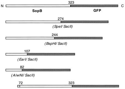
Figure 3.
Illustrations of full-length SopB protein and its truncation derivatives fused to the jellyfish GFP. The number above the SopB and GFP junction in each sketch indicates the particular amino acid residue of SopB at the junction. In the SopB truncation shown at the bottom, the N-terminal 71 amino acid residues of SopB is replaced by a stretch of 14 residues unrelated to SopB.

