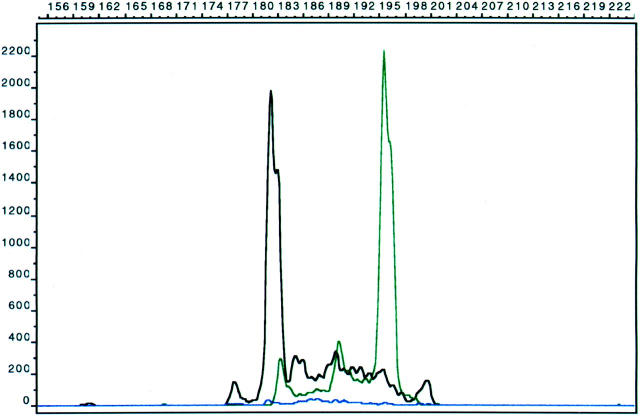
Figure 2.
Capillary electrophoresis method showing biallelic clonal rearrangements with the Jγ1/2 (black) and JγP1/2 (green) primers in a case of peripheral T-cell lymphoma. The heights of the clonal peaks are greater than two times the polyclonal background. x axis, nucleotide length; y axis, intensity of signal.

