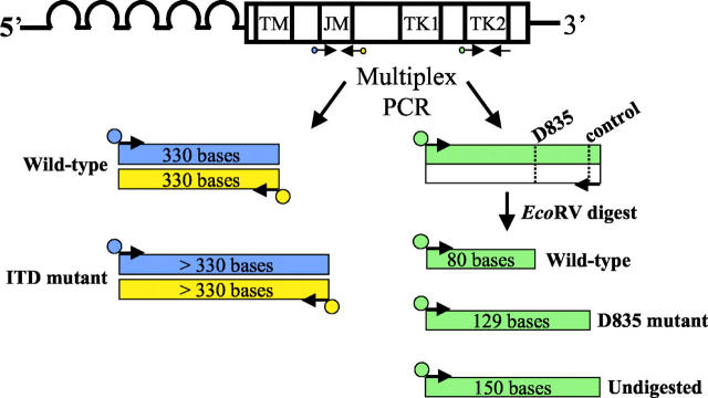
Figure 1.
Diagram of the assay design. The FLT3 gene consists of five extracellular immunoglobulin-like domains, a transmembrane domain (TM), a JM domain, and an interrupted kinase domain (TK1 and TK2). PCR primers flanking the JM domain [forward primer labeled with FAM (blue), reverse primer labeled with NED (yellow)] and primers specific for the TK2 domain [forward labeled with TET (green), reverse unlabeled] are multiplexed into a single PCR reaction. After amplification, the PCR products are digested with EcoRV. The dotted lines in the TK2 PCR product represent the EcoRV cut sites, with the recognition sequence (GATATC). The JM portion of the PCR yields a wild-type PCR product of 330 bases labeled with both FAM and NED. FLT3 ITD mutations result in PCR products that are longer than wild type (>330 bp), also labeled with both FAM and NED. After digestion, the D835 portion of the assay yields wild-type products sizing at 80 bases that are TET labeled. D835 mutant TET-labeled products size at 129 bases, and undigested TET-labeled products size at 150 bases.

