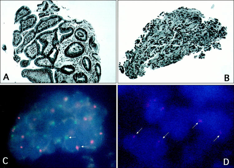
Figure 1.

A: A routine H&E section of an endoscopic gastric biopsy from an elderly male with diffuse gastric mucosal erythema and pebbling, labeled Case A. B: A routine H&E section of a gastric biopsy from a female whose endoscopic examination was highly suggestive of malignant ulcer (contaminant), labeled Case B. C: FISH on tissue section derived from the gastric biopsy illustrated in A. Note the presence of one red signal (X chromosome) and one green signal (Y chromosome) in gastric mucosal cells. D: FISH on the tissue section derived from the gastric biopsy shown in B (contaminant). Note the presence of two red signals (representing X chromosomes) and absence of green signals.
