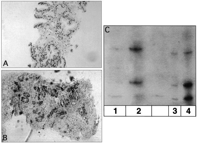
Figure 2.
A: H&E section from Case C composed of benign prostatic epithelium. B: H&E section from Case D showing moderately to poorly differentiated prostatic adenocarcinoma, with Gleason pattern 3 + 5 (Gleason score = 8), and with perineural invasion. C: Autoradiograph of microsatellite analysis of DNA from Case C and Case D (putative source of contaminant). Lane 1: DNA-1 microdissected from the H&E-stained tissue section area of alleged contaminant labeled Case D present on the slide from Case C. Lane 2: DNA-1′ microdissected from tissue sections of a paraffin block of prostate adenocarcinoma from which Case D was suspected to originate. Lane 3: DNA-2 microdissected from the H&E-stained tissue section area of normal prostatic epithelium from Case C. Lane 4: DNA-2′ microdissected from tissue sections originating from a paraffin block of Case C.

