
Figure 1.
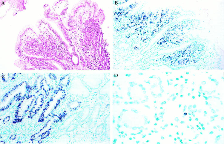
A: H&E stain of invasive gastric adenocarcinoma surrounded by normal surface epithelium. B: EBER in situ hybridization reveals EBER transcripts in the nucleus of the carcinoma cells, but not in the overlying normal surface epithelium, nor in the surrounding benign stromal cells. C: EBER is localized to dysplastic gastric epithelium but not to adjacent normal-appearing glands, implying that EBV infection is an early event in gastric carcinogenesis. D: EBER is localized to the nucleus of a single small lymphoid cell, representing the rare infected lymphocyte that might be found in any previously infected individual. Original magnifications, ×50 (A and B), ×80 (C), and ×150 (D).