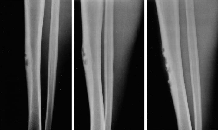
Figure 1.
The Case 2 lesion involving the anterior cortex has multiple radiolucent defects surrounded by areas of cortical thickening and a thick rim of reactive bone at the base. One can observe indolent progression of the radiolucent areas from the first film taken July, 1995 (left) to subsequent ones obtained May, 1996 (center) and December, 1999 (right).

