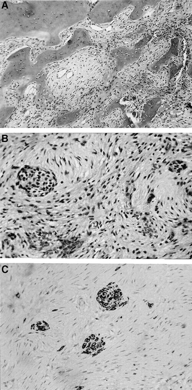
Figure 2.

A: The medullary side of a portion of the reactive cortex is at the top of the left hand corner. It is fairly mature, but lacks well-developed Harversian systems and stress lines. Connected to it and protruding toward the underlying lesion, are thickened trabeculae of irregular, immature bone variably rimmed by osteoblasts. Osteoclastic remodeling can be seen in the lower right hand corner. B: The center of the lesion is characterized by a bland proliferation of cells having spindle shaped, often slender, nuclei arranged in a loose storiform pattern. Embedded within this stroma are two nests of epithelial cells. C: An immunohistochemical preparation for cytokeratins highlights three nests of epithelial cells. In addition, multiple keratin reactive single cells within the stroma, not otherwise detectable, can be appreciated.
