Abstract
Short tandem repeats (STRs) are highly polymorphic DNA sequences in the human genome. STR genotype analysis is used for human identity testing and to monitor bone marrow engraftment after allogeneic transplantation. 1 2 3 Engraftment analysis requires one or more informative STR loci that distinguish recipient from donor. The following case illustrates that chromosome loss in tumor cells during the course of disease may cause corresponding loss of an STR locus. This circumstance is a potential source of error in the interpretation of engraftment analysis, especially if only one informative allele is used to monitor engraftment.
Case Report
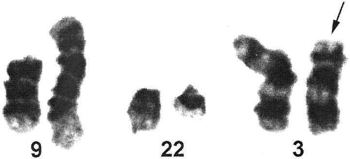
A 48-year-old woman with chronic myeloid leukemia (CML) in chronic phase received an allogeneic peripheral blood stem cell transplant from a related male donor. The post-transplant clinical course was complicated by mild graft-versus-host disease. At 3 months after transplantation, a bone marrow examination showed normocellular trilineage hematopoiesis negative for morphological evidence of CML. Chromosome analysis of the bone marrow showed a normal male karyotype. RT-PCR for BCR/ABL was negative. Three months thereafter, the patient relapsed with CML in blast transformation. A bone marrow examination showed sheets of blasts estimated to account for approximately 80% of the marrow cellularity. Flow cytometry of the blasts showed an immunophenotype (CD45, CD19, CD20, CD10, CD34, HLA-DR) consistent with acute lymphoblastic leukemia, precursor B-cell type. Chromosome analysis showed a female karyotype positive for the Philadelphia chromosome with additional structural and numerical abnormalities including partial deletion of one copy of chromosome (3)(p21) (Figure 1) .
Figure 1.
Karyotype of blasts shows t(9;22)(q34;q11) and partial loss of one copy of chromosome 3p at(3)(p21)(arrow).
Analysis of Engraftment
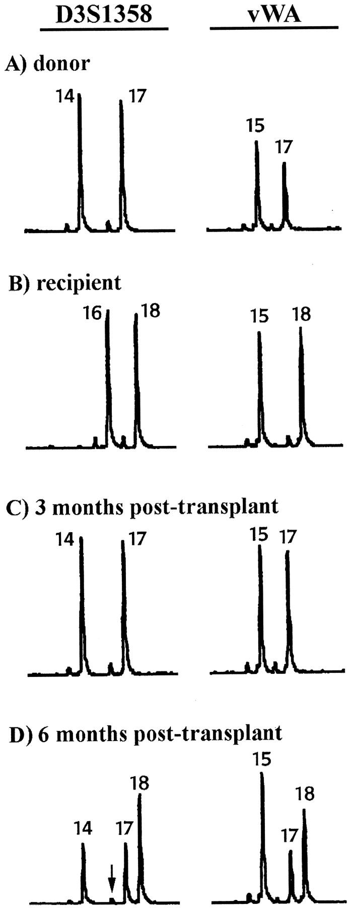
STR analysis of donor and pre-transplant recipient DNA samples from peripheral blood was performed to determine the genotype at three loci (D3S1358, vWA, and FGA) using the AmpFlSTR Blue PCR amplification kit (PE Applied Biosystems, Foster City, CA). 1 After amplification, samples were analyzed on an ABI 373A DNA Sequencer (Applied Biosystems). Fragment size and peak area data were determined by GeneScan and GenoTyper software (Applied Biosystems). 1 The D3S1358 and vWA loci show informative alleles that distinguish donor(Figure 2A) from recipient (Figure 2B) . At the D3S1358 locus, donor and recipient are both heterozygous with no shared alleles. Thus, two informative donor alleles (14 and 17) and two informative recipient alleles (16 and 18) are observed. At the vWA locus, donor and recipient are both heterozygous with one shared allele (15). Thus, one informative donor allele (17) and one informative recipient allele (18) are observed. The genotypes for donor and recipient at the FGA locus (not shown) are identical and therefore uninformative.
Figure 2.

STR genotypes at the D3S1358 and vWA loci for pre- and post-transplant peripheral blood samples. A:Donor. B: Recipient pre-transplant. C: Recipient at 3 months post-transplantation shows genotype identical to the donor indicating 100% donor DNA. D: Recipient at 6 months post-transplantation shows donor and recipient alleles corresponding to a mixture of donor and recipient DNA. Arrow indicates loss of the recipient informative allele D3S1358 (16). Small peak beneath arrow is a stutter peak derived from the donor D3S1358 (17) allele.
Small stutter peaks derived from the main amplified peaks are also present in Figure 2 . Stutter peaks are an artifact of STR PCR amplification that may arise from “slippage” during the PCR process. Stutter peaks of tetranucleotide STRs are 4 bp shorter than the main peak and usually have a peak area close to 5% of the main peak area. 1 In Figure 2A , stutter peaks amplified at the donor D3S1358 locus are present in the (13) position, derived from the D3S1358 (14) allele, and in the (16) position, derived from the D3S1358 (17) allele. Small stutter peaks amplified at the donor vWA locus are present in the (14) position, derived from the vWA (15) allele, and the (16) position, derived from the vWA (17) allele. Similar stutter peaks amplified at the recipient D3S1358 and vWA loci are present in Figure 2B .
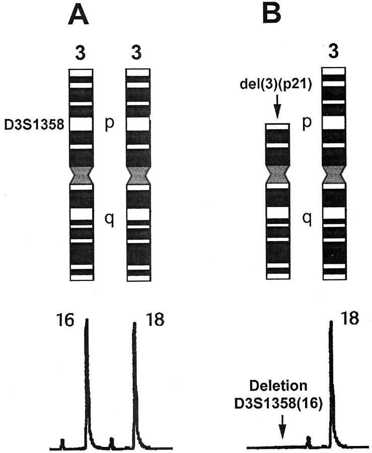
At 3 months post-transplantation, the peripheral blood genotype (Figure 2C) is identical to that of the donor. Quantitative engraftment analysis (see below) yields 100% donor DNA, consistent with full engraftment and disease remission. At 6 months post-transplantation, the peripheral blood genotype (Figure 2D) shows a mixture of donor and recipient alleles, consistent with disease relapse. The vWA locus shows the donor allele (17), the recipient allele (18), and the shared allele (15). Quantitative engraftment analysis at the vWA locus (see below) yields approximately 70% recipient DNA. The D3S1358 locus shows two donor alleles (14 and 17), but only one recipient allele (18). The recipient allele (16) is missing. Only a small stutter peak is present in the (16) position. The D3S1358 locus is located on chromosome (3)(p21). Loss of the recipient allele (16) in DNA from the patient’s leukemic blasts, therefore, corresponds to partial deletion of one copy of chromosome 3p in the karyotype (Figure 3) .
Figure 3.
Correspondence of D3S1358 chromosome locus with STR genotype. The D3S1358 locus is located on the short arm of chromosome 3 at (3)(p21). A: Schematic representation of chromosome 3 pair. The short arm and long arm of chromosome 3 are indicated by the letters p and q, respectively. Heterozygous STR genotype is shown beneath the corresponding chromosomes. One copy of chromosome 3 has the D3S1358 (16) allele. One copy of chromosome 3 has the D3S1358 (18) allele. B: Partial deletion of one copy of chromosome 3p at (3)(p21) corresponds to loss of the D3S1358 (16) allele.
Calculation of Recipient and Donor DNA Percentages
The percentage of donor and recipient DNA can be quantitated from STR analysis to estimate the degree of bone marrow engraftment. 1 Ratios of the peak areas of donor and recipient alleles at an informative locus are used to calculate the DNA percentages in post-transplant samples. Specific formulas for calculating the percentage of donor or recipient DNA are based on whether the informative loci are heterozygous or homozygous and whether the donor and recipient share any alleles. Because recipient DNA is usually the minor component in post-transplant samples, calculation of the percentage of recipient DNA is usually performed. The percentage of donor DNA is then derived by subtracting the percentage of recipient DNA from 100%.
At the vWA locus in Figure 2D , the following formula calculates the percentage of recipient DNA, where
 |
;
 |
.
 |
At the D3S1358 locus in Figure 2D , the following formula would be used to calculate the percentage of recipient DNA, where A(16) = the area of the peak of recipient allele (16); A(18) = the area of the peak of recipient allele (18); A(14) = the area of the peak of donor allele (14);
 |
.
 |
However, due to chromosomal deletion, the D3S1358 (16) allele peak is missing from the STR genotype in Figure 2D . Only a small stutter peak is present. Calculation of the percentage of recipient DNA from the formula would therefore lead to an erroneous result.
Discussion
Malignant cells may exhibit genetic instability resulting in partial or complete loss of chromosomes. Chromosome loss is associated with loss of corresponding genetic loci. Hematological malignancies often show karyotypes with multiple structural and numerical abnormalities, especially after treatment with chemotherapy and/or radiation. 4 As illustrated in this case report, loss of an STR allele corresponding to a specific chromosome loss can occur in leukemia relapse after transplantation. We have observed two separate incidences of STR allele loss in our analysis of post-transplant samples from approximately 140 transplant recipients with hematological malignancy. Loss of an informative allele is important as a potential source of error in engraftment analysis. 5
STR genotype analysis of bone marrow transplant engraftment requires the identification of informative STR loci that distinguish recipient DNA from donor DNA. In theory, a single informative recipient allele within a single locus is sufficient to monitor engraftment after transplantation. Our case illustrates that a single informative allele would not be sufficient for engraftment analysis if loss of the allele occurs due to genomic instability. For example, if the deleted allele D3S1358 (16) had been the only informative recipient allele in our CML case, then the patient’s leukemic blood at relapse would have been interpreted incorrectly as containing 100% donor DNA. To avoid possible errors in engraftment analysis, we recommend that two or more informative STR alleles, preferably at different loci, be used to monitor engraftment.
This case study also illustrates the importance of correlating engraftment analysis with available clinical and laboratory data. Hematopathology, flow cytometry, and cytogenetics results may be helpful in interpreting complex or confusing engraftment data. We recommend confirming that the engraftment results are consistent with the hematopathology report and other laboratory studies, where available, to avoid possible error and to assure quality.
Acknowledgments
We thank Jeff Bowen of the CAVHS Medical Media Service for his kind assistance in graphic design.
Address reprint requests to Dr. S.A. Schichman, Pathology and Laboratory Medicine Service (113/LR), Central Arkansas Veterans Healthcare System, 4300 West 7th Street, Little Rock, AR 72205. E-mail: schichman@exchange.UAMS.edu.
References
- 1.Schichman SA, Suess P, Vertino AM, Gray PS: Comparison of short tandem repeat and variable number tandem repeat genetic markers for quantitative determination of allogeneic bone marrow transplant engraftment. Bone Marrow Transplant 2002, 29:243-248 [DOI] [PubMed] [Google Scholar]
- 2.Van Deerlin VM, Leonard DG: Bone marrow engraftment analysis after allogeneic bone marrow transplantation. Clin Lab Med 2000, 20:197-225 [PubMed] [Google Scholar]
- 3.Nuckols JD, Rasheed BK, McGlennen RC, Bigner SH, Stenzel TT: Evaluation of an automated technique for assessment of marrow engraftment after allogeneic bone marrow transplantation using a commercially available kit. Am J Clin Pathol 2000, 113:135-140 [DOI] [PubMed] [Google Scholar]
- 4.Heim S, Mitelman F: Cancer Cytogenetics ed 2. 1995:33-309 Wiley-Liss New York
- 5.Zhou M, Sheldon S, Akel N, Killeen AA: Chromosomal aneuploidy in leukemic blast crisis: a potential source of error in interpretation of bone marrow engraftment analysis by VNTR amplification. Mol Diagn 1999, 4:153-157 [DOI] [PubMed] [Google Scholar]


