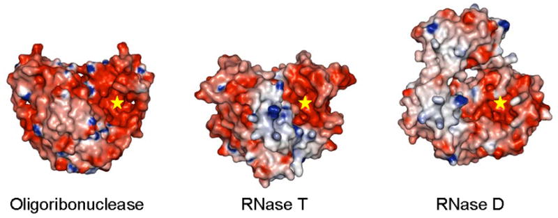
Figure 6.

Comparison of the structures of the three DEDD family exoribonucleases from E. coli. Molecular surfaces colored by electrostatic potential are shown for these enzymes with one DEDD domain from each aligned in the same orientation. The active centers of these enzymes are highlighted (“star”).
