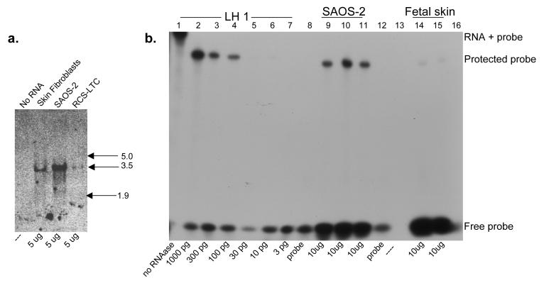
Figure 6.
a: Northern Blot analysis of LH1 mRNA in SAOS-2 cells and skin
5 μg of SAOS-2, human fetal skin and RCS-LTC chondrocyte cell line total RNA was resolved on a 1.2% agarose-formaldehyde gel, blotted to nitrocellulose membrane, probed with a 32P-labelled LH1 cDNA, and detected by autoradiography. RNA kilobase markers are shown at right.
b: Quantitation of LH1 mRNA in SAOS-2 cells and skin by RNAase protection.
RNA was hybridized to a 32P-labelled LH1 antisense probe, digested with nuclease to remove the non-homologous portion of the probe, resolved on a denaturing 6% acrylamide gel, and detected by autoradiography. Lane 1, Full-length probe prior to nuclease digestion. Lanes 2-7, Control LH1 RNA reactions containing 1000, 300, 100, 30, 10, and 3 pg LH1 RNA respectively. Lanes 9-11, Replicate reactions containing 10 μg SAOS-2 RNA. Lanes 14 and 15, Replicate reactions containing 10 μg human fetal skin RNA. Lane 8 and12, Probe only. Lanes 13 and 16 are empty.

