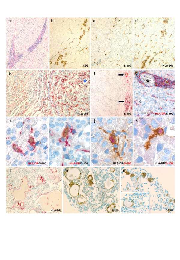
Figure 1.
Microphotographs from Case 3 (a through d) to illustrate basic pattern of inflammatory reaction common to all three adenomas in this report: i.e., angiocentric infiltrates of T lymphocytes (b); colonization of tumor interstitium by arborized cells immunoreactive for S-100 protein (c) and HLA-DR (d), the respective staining patterns of which are felt to be largely overlapping. As exemplified by index Case 1 (e), dendritic cell reaction selectively involves the adenoma compartment (*), while only native fibrohistiocytic elements of peritumoral pituitary stain for HLA-DR. Lesional interface is highlighted by dotted line. Conventional gonadotroph cell adenoma from the control series (f) is devoid of S-100+ dendritic cells. Arrows point to activated folliculo-stellate cells in adjacent nontumorous acini to technically validate staining reaction. Overview of double immunostained inflammatory focus (g) reveals simultaneous positivity for S-100 protein and HLA-DR in most arborized cells both around intratumoral vessel (*) and within interstitium (arrows). Stellate morphology of HLA-DR/S-100 coexpressing cells is appreciated both along vascular sleeves (h and j) of tumoral microcirculation and as these tend to intimately commingle with adenoma cells (i and k). In order for reproducibility of specific labeling to be ensured, immunoreactions for both epitopes have been duplicated while interverting chromogens. Unique to the prolactin cell adenoma in Case 2 is the finding of adenomatous follicles (l and m) lined in part by HLA-DR immunoreactive FSCs. While rare follicles with nearly identical morphology also did stain for GFAP (n), actual coexpression could not be demonstrated. Immunohistochemical specimens depicted in b – d, and m – n were developed with horseradish peroxidase and 3,3'-diaminobenzidine; slides shown in e – f, and l are visualized using streptavidin-biotin-complex/alkaline phosphatase and new fuchsin-naphtol AS-BI as chromogen. In the double labeling studies (g through k), antigen names typeset in either red or black indicate fuchsin-naphtol and diaminobenzidine, respectively. Original magnification: a, l through n – ×200; b through f – ×100; g – ×400; h through k – ×1000 (oil immersion).

