
Figure 9. PERK Inhibition Results in Increased Laminin-5 Production, Secretion and Deposition.
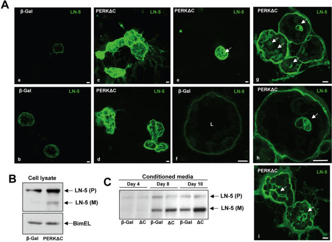
(A) Confocal images showing the equatorial cross sections through β-Gal (A-a, A-b and A-f) and PERKΔC (A-c to A-e and A-g to A-i) acini stained for Laminin-5 (green), on Day 8 in 3D. Note the increased staining and disorganized Laminin-5 deposition in PERKΔC cells as compared to proper basal localization in the β-Gal control cells. Panels A-e and A-g through A-i (arrows) show details of intra-acinar deposition of Laminin-5 within and around cells in the filled lumen. Scale bars = 10 µm; A-h is a magnified image of an acinus shown in A-g. (B) Western blot analysis from β-Gal and PERKΔC cell lysates shows the precursor (P) and mature (M) forms of LN-5. BimEL was used as loading control. (C) Western blot for Laminin-5 secreted into the conditioned media by β-Gal and PERKΔC cells grown in 3D-Matrigel. For conditioned media, samples corresponding to days 4 and 8, the supernatant was collected after 96 hrs of 3D culture. For the conditioned media sample corresponding to day 10, the supernatant was collected after 48 hrs of 3D culture. ΔC = MCF10A-PERKΔC cells. Note that while in cell lysates (B) the precursor is predominantly detected, in the conditioned media there is stronger detection of the mature form of the Laminin-5-γ2 subunit.