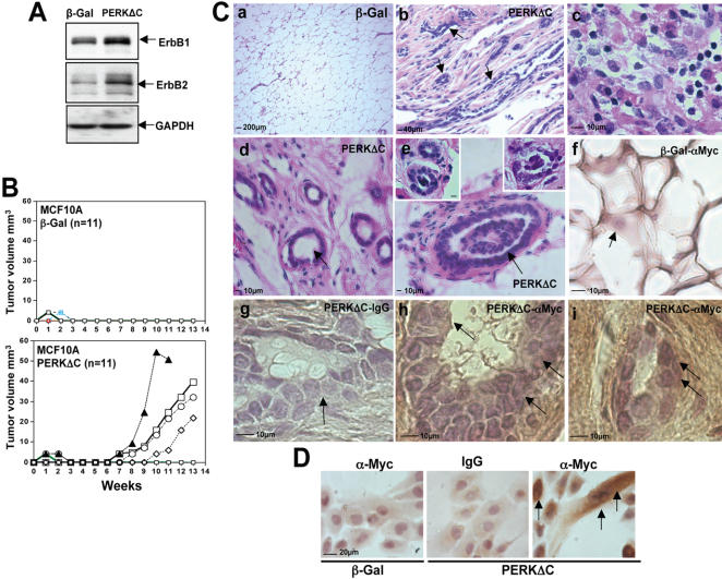
Figure 10. PERK Inhibition in MCF10A Cells Favors Mammary Tumor Formation.
(A) Western blot for ErbB1 (EGFR) and ErbB2 (Her2) levels in adhered β-Gal or PERKΔC cells show increased levels when PERK is inhibited. GAPDH was used as loading control. (B) β-Gal or PERKΔC cells were injected orthotopically into the contralateral abdominal mammary fat pads of 3 week-old female nude mice (see methods for details). Post-implantation mice were monitored biweekly for tumor take and when detected tumor diameters were measured and the volume was calculated and plotted as described in methods. Note that none of the mice implanted with β-Gal-MCF10A cells developed tumor nodules. (C) Histology of mammary glands and tumors in mice implanted with β-Gal and PERKΔC expressing MCF10A cells. (C-a) H&E staining of a cleared mammary fat pad inoculated with β-Gal cells. Only adipose tissue and occasional stromal cells that were negative for β-galactosidase activity was observed. (C-b) H&E staining of a PERKΔC tumor lesion. Note the disorganization of the fibrotic-epithelial tissue. Arrows depict the presence of epithelial cells forming acinar or duct-like structures within the PERKΔC tumor lesion. (C-c) Higher magnification of intratumoral accumulations of cells without a defined architecture but comprised of a mixture of epithelial (larger nuclei and pink cytoplasm) and inflammatory cells (smaller darkly stained nuclei). (C-d and e) PERKΔC cells form acini-like structures with an empty lumen or show hyperplastic growth and a repopulated lumen (C-e, top left and right insets). (C-f) Anti-Myc (αMyc) staining in control β-Gal injected mammary fat pads. Note that only a light background signal is observed in adipocytes. (C-g) Histological section of a PERKΔC acinus-like structure stained with a non-specific IgG (arrow denotes the lack of staining in these epithelial cells) or with anti-Myc 9E10 mAb (C-h and C-i); arrows denote the brown staining generated by Myc-tag detection. (D) β-Gal (left panel) or PERKΔC (middle and right panels) cells grown on 2D coverslips and fixed and stained with a non-specific IgG (IgG) or with an anti-Myc 9E10 mAb (α-Myc). The Myc staining was characteristic of intracellular cistern distribution.

