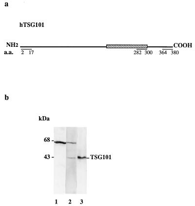
Figure 1.
Generation and analysis of anti-TSG101 antibody. (a) Map of TSG101 protein-coding region showing the location of the N-terminal (amino acids 2–17), coiled-coil domain (amino acids 282–300), and C-terminal peptides (amino acids 364–380), which are identical or highly conserved in human and mouse and were used as antigens. Peptide locations are designated according to the TSG101 sequence shown in ref. 2. (b) Polyclonal antibody detecting a band of 43 kDa in mammalian extract (lane 2); analysis using prebleed serum from the same rabbit (lane 1). Lane 3 shows affinity-purified antibody against the TSG101 coiled-coil domain detecting a single band, migrating at the same location as the mammalian signal, in an extract of Sf9 cells overexpressing TSG101. Purified antibodies generated against the N-terminal or C-terminal domains of TSG101 detected the same band.

