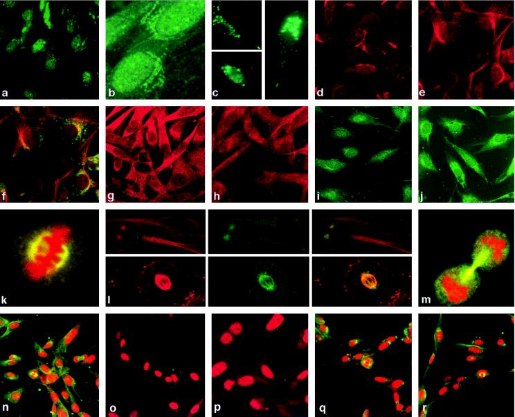
Figure 2.
Cell cycle-dependent localization of TSG101. Cells were synchronized at G0 (a), G1 (b), G1/S (i), and S (j) stages of the cell cycle and stained with affinity-purified anti-TSG101 antibody. GFP-TSG101 fusion protein localization in cycling cells: c Lower, nuclear staining; c Upper, diffuse cytoplasmic staining; c Right, asymmetric cytoplasmic staining. Cells stained with the Golgi-specific marker β-COP (d) and TSG101 (e). Cells costained with anti-TSG101 antibody (red) and Bodipy FL C5-ceramide (green) showing colocalization (yellow) (f) Brefeldin A-treated cells stained for β-COP (g) and TSG101 (h) showing cytoplasmic dispersion. Mitotic cells (k) showing staining of TSG101 (green) in mitotic spindle and propidium iodide-stained metaphase chromatin (red). (l Upper × 3) Localization of tubulin (red), TSG101 (green), and both (yellow) in centrosomes. (l Lower × 3) The staining of a mitotic spindle, as above. (m) Staining of TSG101 in midbody (green); condensed chromatin is stained in red by propidium iodide. Cells stained with propidium iodide (red) and purified anti-TSG101 antibody (green) (n), and with the same antibody preincubated with the antigen peptide (o) or two irrelevant TSG101 peptides (q and r), are compared with cells stained with prebleed from the same rabbit purified in the same fashion as the immune sera (p). All photographs except for GFP-fusion study were made by using confocal microscopy.

